Bone Imaging Suite
The Bone Imaging Suite in the Clinical Research Facility at the Northern General Hospital has a team of dedicated scan technicians who have undergone formal training and accreditation from the National Osteoporosis Society.

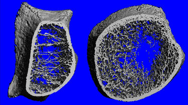
The structural and geometric properties of bone can be studied in man using state-of-the-art equipment purchased with the support of the Medical Research Council and the National Institute of Health Research. This suite is based in the at the Northern General Hospital.
The CRF provides a welcoming reception, with a a waiting room and private data rooms, consulting suites and treatment rooms, bed bays and single rooms, and an investigation laboratory. All staff are trained and operate to Good Clinical Practice standards, and our clinical trials nursing team ensures that volunteers receive personalised care throughout their visits.
The specialised research Bone Imaging Suite consists of the following devices:
- Horizon A DXA scanner (Hologic Inc.)
- XtremeCT HR-pQCT scanner (Scanco Medical AG)
The purchase of the XtremeCT HR-pQCT scanner was made possible through the National Institute for Health and Care Research (NIHR).
The purchase of the Horizon A DXA scanner was kindly funded through the Louise Victoria Craddock Memorial Fund, Weston Park Hospital Cancer Charity.
For further information, please contact Bone Imaging Lead, Dr Margaret Paggiosi.
