
Pulmonary imaging
Our research focus is the application of magnetic resonance imaging (MRI) techniques to pulmonary diseases, with an overarching aim of developing novel imaging biomarkers that can be used to better understand the pathophysiology of lung disease.

91ÖąēĨ has pioneered the use of HP gas MRI over the last 20 years, and is a world leader in the field. In 2015 our centre was licensed for the manufacture of hyperpolarised 3He and 129Xe gas for clinical lung investigations, a world first for this technology.
This work has expanded significantly over recent years with the advent of a new imaging laboratory and now attracts clinical referrals from respiratory physicians across the UK.
Whilst inexpensive and simple to perform, spirometry is insufficiently sensitive to detect early functional changes associated with lung disease. More sensitive methods are needed to allow for early therapy and better patient management, and so we focus on applying hyperpolarised gas MRI to study lung microstructure, and ventilation and gas exchange function; high resolution structural MRI to visualise the lung macrostructure; and dynamic contrast enhanced MRI to assess lung perfusion. These methods can be applied to assess lung structure and function in a range of pulmonary disorders.
Research into these techniques is ongoing and areas of interest include:
- Chronic obstructive pulmonary disease (COPD)
- Asthma
- Interstitial lung disease (ILD), including Idiopathic Pulmonary Fibrosis (IPF)
- Cystic fibrosis (CF)
- Pulmonary vascular disease
- Pre-term birth-related lung disease and primary ciliary dyskinesia (PCD)
Overarching projects in clinical translation:
- . NIHR - Research Professorship. PI: Wild
- . MRC - Resources and infrastructure Research Grant. PI: Wild
COPD and Asthma
Asthma and COPD are among the most common respiratory diseases, with COPD being the third leading cause of death worldwide. We use hyperpolarised gas MRI to visualise the distribution of ventilation in the lung and obtain imaging biomarkers of ventilation that show increased sensitivity over spirometry in COPD and asthma. Diffusion imaging can be used to measure enlarged alveolar airspaces in emphysema, and dynamic contrast enhanced perfusion imaging can visualise the distribution of blood perfusion in the lung microvasculature. In patients with severe COPD, time-resolved ventilation imaging can detect collateral ventilation, the key factor that determines the success or failure of lung volume reduction surgery (LVRS) using endobronchial valves.
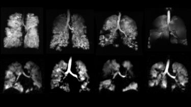
Hyperpolarised gas MRI does not involve ionising radiation exposure and therefore can be performed repeatedly; e.g. multiple times in a single day to assess treatment response to bronchodilator therapy, or over longer time-periods to evaluate longer-term therapies. We have carried out studies to assess treatment response to novel drugs in patients suffering from these diseases. Furthermore, we have obtained Medicines and Healthcare products Regulatory Agency (MHRA) approval to perform these scans as part of routine , to aid clinicians in managing those patients with challenging diagnoses. Our clinician collaborators report that visualising regional lung ventilation provides a greater clinical insight than spirometry alone.

Projects, Collaborators & Publications
- Current Projects / Grants
-
- ADPro - Observational study of obstructive lung disease in patients with COPD and asthma (funded by AstraZeneca)
- Past Projects / Grants
-
- Study to assess the effect of Indacaterol/Glycopyrronium on ventilation and perfusion in COPD (funded by Novartis),
- - Airway Disease Predicting Outcomes through Patient Specific Computational Modelling
- Fevipripant asthma study with the University of Leicester (funded by EU FP7 Air PROM and Novartis)
- Investigation of the use of MRI to detect changes in ventilation-perfusion relationships in COPD (funded by GSK)
- Collaborators
-
- (University of 91ÖąēĨ)
- Ian Sabroe (91ÖąēĨ Teaching Hospitals)
- and (University of Leicester)
- Rod Hughes (AstraZeneca) and (University of Manchester)
- Key Publications
-
- . Chan HF, Collier GJ, Weatherley ND, Wild JM. Magn Reson Med. 2019 May;81(5):2959-2971. doi: 10.1002/mrm.27608.
- . Stewart NJ, Chan HF, Hughes PJC, Horn FC, Norquay G, Rao M, Yates DP, Ireland RH, Hatton MQ, Tahir BA, Ford P, Swift AJ, Lawson R, Marshall H, Collier GJ, Wild JM. J Magn Reson Imaging. 2018 Mar 5;48(3):632-42.
- . Marshall H, Collier GJ, Johns CS, Chan HF, Norquay G, Lawson RA, Wild JM. J Magn Reson Imaging. 2019 Apr;49(4):1195-1197. doi: 10.1002/jmri.26273.
- . Horn FC, Marshall H, Collier GJ, Kay R, Siddiqui S, Brightling CE, Parra-Robles J, Wild JM. Radiology. 2017 Sep;284(3):854-861. doi: 10.1148/radiol.2017160532.
- . Marshall H, Deppe MH, Parra-Robles J, Hillis S, Billings CG, Rajaram S, Swift A, Miller SR, Watson JH, Wolber J, Lipson DA, Lawson R, Wild JM. Thorax. 2012 Jul;67(7):613-7. doi: 10.1136/thoraxjnl-2011-200864.
Interstitial lung disease
Interstitial lung disease (ILD) covers a wide range of pulmonary disorders, including idiopathic pulmonary fibrosis (IPF) and hypersensitivity pneumonitis (HP), which can present as progressive fibrosis (scarring) of the lung tissue. ILD, whether fibrotic or not, is typically characterised by gas exchange functional impairment in the lung. The imaging methods that we use to help better assess the progression of disease in these patients include: diffusion-weighted MRI with hyperpolarised 3He or 129Xe, MR spectroscopy / spectroscopic imaging of 129Xe dissolved in the lung tissue and blood, and 1H dynamic contrast-enhanced (DCE) MRI. DW-MRI provides information on the microstructure of the alveoli, while 129Xe spectroscopy / spectroscopic imaging measures provide information on fibrosis and tissue thickening, and their influence on gas exchange. DCE MRI provides information on the passage of blood through the lung and its change over time in these patients. In addition, through recent collaborations with Wisconsin and the Mayo Clinic, we have implemented a high-resolution UTE MRI sequence for macrostructural assessment of the lung of comparable quality to CT, and have investigated quantitative CT biomarkers, in patients with ILD.


We are using high resolution UTE MR images to study patients with a number of subtypes of interstitial lung disease (ILD) as part of the , and we are able to detect changes similar to those seen on CT.
Projects, Collaborators & Publications
- Current Projects / Grants
-
- Tristan:
- Investigation into prognostic indicators of Idiopathic Pulmonary Fibrosis using structural-functional pulmonary MRI assessment (funded by Boehringer Ingelheim)
- Assessment of lung function in patients with idiopathic pulmonary fibrosis using hyperpolarised xenon MRI, a sub-study of the (funded by Galapagos)
- Collaborators
-
- (University of 91ÖąēĨ)
- Steve Renshaw (University of 91ÖąēĨ)
- Nazia Chaudhuri (University of Manchester NHS Foundation Trust)
- Paul Ford ()
- & Ronald Karwoski (Mayo Clinic; )
- (University of Wisconsin)
- (University College London, CALIPER analysis)
- (Iowa, AMFM quantitative ILD CT software)
- Key Publications
-
- . Saunders LC, Eaden JA, Bianchi SM, Swift AJ, Wild JM. Magn Reson Med. 2020 Dec;84(6):3088-3102.
- . Weatherley ND, Stewart NJ, Chan HF, Austin M, Smith LJ, Collier G, Rao M, Marshall H, Norquay G, Renshaw SA, Bianchi SM, Wild JM. Thorax. 2019 May;74(5):500-502.
- . Chan HF, Weatherley ND, Johns CS, Stewart NJ, Collier GJ, Bianchi SM, Wild JM. Radiology. 2019 Apr;291(1):223-229.
- . Stewart NJ, Leung G, Norquay G, Marshall H, Parra-Robles J, Murphy PS, Schulte RF, Elliot C, Condliffe R, Griffiths PD, Kiely DG, Whyte MK, Wolber J, Wild JM. Magn Reson Med. 2015 Jul;74(1):196-207.
Cystic fibrosis
One key issue in monitoring patients with CF is the global information provided from lung function, rather than the locality and regional extent of lung disease, which can only be accessed with imaging. CT is often used clinically when managing patients with CF, however repeated exposure to ionising radiation, particularly in children, is a concern and has led our centre and others to pursue MRI for lung functional assessment of patients with CF.
We were the first centre to perform paediatric studies in CF, and have applied hyperpolarised gas and structural UTE MRI in CF in a number of studies, scanning patients as young as 6 years old. These imaging methods can detect subclinical changes in lung function and involve no ionising radiation, so are well-suited to longitudinal follow-up / monitoring of disease in these patients.

We are also involved in a number of collaborative studies investigating structure-function relationships in CF and evaluating novel proton MRI methods developed by our partners.

Projects, Collaborators & Publications
- Current Projects / Grants
-
- MMAVIC study - MRI & multiple breath washout to assess ventilation in cystic fibrosis
- - Validation of bi- and three-dimensional Fourier Decomposition to assess lung ventilation and perfusion compared to CT, hyperpolarized gases and contrast-enhanced MRI
- Collaborators
-
- Chris Taylor (91ÖąēĨ Childrenâs Hospital)
- (91ÖąēĨ Childrenâs Hospital)
- (University of Manchester)
- (Erasmus MC, Rotterdam)
- (Hannover Medical School)
- Key Publications
-
- . Smith LJ, Marshall H, Bray J, Wildman M, West N, Horsley A, Wild JM. J Cyst Fibros. 2021 Jul;20(4):625-631.
- . Smith LJ, Horsley A, Bray J, Hughes PJC, Biancardi A, Norquay G, Wildman M, West N, Marshall H, Wild JM. Eur Respir J. 2020 Jul 6:2000441.
- . Smith LJ, Collier GJ, Marshall H, Hughes PJC, Biancardi AM, Wildman M, Aldag I, West N, Horsley A, Wild JM. Eur Respir J. 2018 Nov 8;52(5):1800821.
- . Smith L, Marshall H, Aldag I, Horn F, Collier G, Hughes D, West N, Horsley A, Taylor CJ, Wild J. Am J Respir Crit Care Med. 2018 Feb 1;197(3):397-400.
- . Marshall H, Horsley A, Taylor CJ, Smith L, Hughes D, Horn FC, Swift AJ, Parra-Robles J, Hughes PJ, Norquay G, Stewart NJ, Collier GJ, Teare D, Cunningham S, Aldag I, Wild JM. Thorax. 2017 Aug;72(8):760-762.
- . Woodhouse N, Wild JM, van Beek EJ, Hoggard N, Barker N, Taylor CJ. J Magn Reson Imaging. 2009 Nov;30(5):981-8.
- . van Beek EJ, Hill C, Woodhouse N, Fichele S, Fleming S, Howe B, Bott S, Wild JM, Taylor CJ. Eur Radiol. 2007 Apr;17(4):1018-24.
- . McMahon CJ, Dodd JD, Hill C, Woodhouse N, Wild JM, Fichele S, Gallagher CG, Skehan SJ, van Beek EJ, Masterson JB. Eur Radiol. 2006 Nov;16(11):2483-90.
- . Koumellis P, van Beek EJ, Woodhouse N, Fichele S, Swift AJ, Paley MN, Hill C, Taylor CJ, Wild JM. J Magn Reson Imaging. 2005 Sep;22(3):420-6.
Pulmonary vascular disease
Pulmonary vascular disease is diagnosed using invasive techniques such as right heart catheter or nuclear medicine imaging. Our research focus is the use of MRI for non-invasive diagnostic and prognostic markers in pulmonary vascular disease. We have developed image processing techniques and software to allow for analysis of quantitative metrics of cardiopulmonary function, such as pulmonary VQ matching and T1 relaxation time.
See also: Cardiovascular Imaging


Projects, Collaborators & Publications
- Current Projects / Grants
-
- . Wellcome Trust Clinical Research Career Development Fellowship â PI: Swift
- Collaborators
-
- David Kiely (University of 91ÖąēĨ)
- Roger Thompson (University of 91ÖąēĨ)
- (University of 91ÖąēĨ)
- Robin Condliffe (University of 91ÖąēĨ)
- Allan Lawrie (University of 91ÖąēĨ)
- (Hannover Medical School)
- Key Publications
-
- . Johns CS, Swift AJ, Rajaram S, Hughes PJC, Capener DJ, Kiely DG, Wild JM. J Magn Reson Imaging. 2017 Dec;46(6):1693-1697.
- . Swift AJ, Capener D, Johns C, Hamilton N, Rothman A, Elliot C, Condliffe R, Charalampopoulos A, Rajaram S, Lawrie A, Campbell MJ, Wild JM, Kiely DG. Am J Respir Crit Care Med. 2017 Jul 15;196(2):228-239.
- . Marshall H, Kiely DG, Parra-Robles J, Capener D, Deppe MH, van Beek EJ, Swift AJ, Rajaram S, Hurdman J, Condliffe R, Elliot CA, Wild JM. Am J Respir Crit Care Med. 2014 Sep 1;190(5):e18-9.
- . Rajaram S, Swift AJ, Telfer A, Hurdman J, Marshall H, Lorenz E, Capener D, Davies C, Hill C, Elliot C, Condliffe R, Wild JM, Kiely DG. Thorax. 2013 Jul;68(7):677-8.
- . Rajaram S, Swift AJ, Capener D, Telfer A, Davies C, Hill C, Condliffe R, Elliot C, Hurdman J, Kiely DG, Wild JM. Radiology. 2012 May;263(2):569-77.
Pre-term Birth Related Lung Disease and PCD
In addition to our extensive research in to paediatric CF, we have utilised hyperpolarised gas MRI to study the lung ventilation and microstructural changes associated with pre-term birth related lung disease () and congenital diaphragmatic hernia (CDH).

In a study using ventilation MRI in children with primary ciliary dyskinesia (PCD), we found ventilation defects even in the presence of normal lung clearance index and FEV1, suggesting that ventilation MRI is a sensitive method for detecting lung disease in children with PCD.

Projects, Collaborators & Publications
- Current Projects / Grants
-
- ERS/EU Marie Curie Fellowship
- Collaborators
-
- Porus Bustani (91ÖąēĨ Teaching Hospitals)
- (Cardiff University)
- and ()
- Marjolein Spoel & (Erasmus MC, Rotterdam)
- Key Publications
-
- . Smith LJ, West N, Hughes D, Marshall H, Johns CS, Stewart NJ, Chan HF, Rao M, Capener DJ, Bray J, Collier GJ, Hughes PJC, Norquay G, Schofield L, Chetcuti P, Moya E, Wild JM. Ann Am Thorac Soc. 2018 Dec;15(12):1487-1490.
- . Spoel M, Marshall H, IJsselstijn H, Parra-Robles J, van der Wiel E, Swift AJ, Rajaram S, Tibboel D, Tiddens HA, Wild JM. Pediatr Pulmonol. 2016 May;51(5):517-24. doi: 10.1002/ppul.23325.
People


- Smitha Rajaram (Consultant Radiologist)