Our is housed at the Sorby Centre for Electron Microscopy here at the University of 91Ö±²¥. It was installed with the NanoMEGAS to improve the capabilities we offer.
Dr Jiahui Qi, Experimental Officer - Electron Microscopies: TEM/FIB, has compiled this case study to explain more about what NanoMEGAS can allow our researchers achieve.
Challenges Before NanoMEGAS:
Prior to acquiring the NanoMEGAS system, we encountered limitations in quantitatively interpreting the nanoscale structural changes that occurred during tribological processes. While conventional methods like transmission electron microscopy (TEM) and electron backscatter diffraction (EBSD) offered some insight, they could not fully resolve the fine details of highly deformed structures at the nano-level. Specifically, the team struggled to quantify dislocation densities and detect subtle phase transformations that influence friction and wear. This lack of precision inhibited their ability to predict and improve material performance, particularly for biomedical implants where understanding the interplay between wear, corrosion, and protein interaction is crucial.
Why NanoMEGAS?
- Precession Electron Diffraction (PED): NanoMEGAS provided a breakthrough with its precession electron diffraction technology, offering spatial resolution between 0.5–5 nm and an angular resolution of approximately 0.1°. This capability allowed us to analyse the ultrafine, nanocrystalline structures formed under severe tribological conditions, which were previously very challenging using conventional techniques.
- Resolution of Complex Structures: The ultrafine grain structures that develop in materials like Ti-6Al-4V and CoCrMo during tribocorrosion were difficult to characterise using traditional methods. PED allowed the lab to accurately index different phases and calculate dislocation densities, providing a more comprehensive understanding of the deformation mechanisms at play.
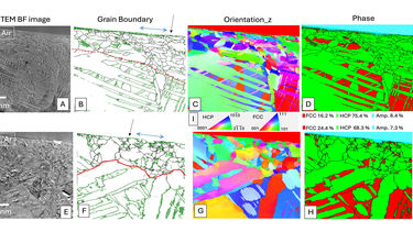
- ASTAR Software Integration: NanoMEGAS's ASTAR software enabled automated phase and orientation mapping, which was crucial for the lab to interpret the highly deformed structures efficiently. This software allowed the lab to correlate the nanoscale structural data with the wear performance of materials.
- Ability to Quantify GND Density: NanoMEGAS’s capabilities to obtain data are then used for calculating the geometrically necessary dislocation (GND) density. This helped the lab evaluate the degree of deformation in different phases and under various tribological conditions. This allowed for more precise predictions regarding how materials will perform under wear and corrosion.
How NanoMEGAS Transformed Our Research:
In one of the research projects, NanoMEGAS played a vital role in understanding the microstructural evolution of Ti-6Al-4V and CoCrMo alloys during tribocorrosion tests conducted in a simulated body fluid environment. Tribocorrosion—an interaction between wear, friction, and electrochemical corrosion—has a significant impact on the performance of biomedical implants. Using NanoMEGAS, we were able to calculate the geometrically necessary dislocation (GND) density and characterize the deformation of tribofilms and worn surface. The ability to measure deformation structures quantitatively at nanoscale precision was critical to uncovering how these tribofilms influence material wear resistance.
Case Study Example: Tribocorrosion of Ti-6Al-4V
The study investigated the tribocorrosion behaviour of Ti-6Al-4V, a commonly used biomedical implant material, in a simulated body fluid environment containing proteins (BSA). The research revealed that the formation of tribofilms significantly reduced friction and wear under certain electrochemical conditions, particularly at positive potentials. NanoMEGAS was used to analyse the microstructure of the worn surface, where it detected the presence of onion-like carbon structures (nanocarbon onions) within the wear debris. These findings suggested that proteins in the fluid contributed to the formation of a protective tribofilm, which improved the implant’s wear performance ​().
Results and Key Findings:
NanoMEGAS enabled the team to achieve several key results that were unattainable with conventional techniques:
- Quantified GND density and identified nanostructural changes under different tribocorrosion conditions.
- Detected the formation of a protective tribofilm that reduced friction and wear, particularly under conditions with a positive potential and protein interaction.
Conclusion:
NanoMEGAS has proven to be an indispensable tool in advancing our understanding of tribological processes at the nanoscale. Its ability to precisely characterise microstructural changes has provided invaluable insights into material performance in a wide range of applications.
For more information or collaboration inquiries, please contact the Sorby Centre for Electron Microscopy at i.ross@sheffield.ac.uk or visit 91Ö±²¥ Electron Microscopy Facility.
List of publications:
1. Qi J, Cole T, Foster A & Rainforth WM (2024) . Wear, 556-557, 205523-205523.
2. Qi J, Ma L, Gong P & Rainforth WM (2023) . Wear, 518-519, 204649-204649.
3. Qi J, Guan D, Nutter J, Wang B & Rainforth WM (2022) . Acta Biomaterialia, 141, 466-480.
4. Xu Y, Qi J, Nutter J, Sharp J, Bai M, Ma L & Rainforth WM (2021) . Tribology International, 163, 107147-107147.
5. Namus R, Nutter J, Qi J & Rainforth WM (2021) . Tribology International, 160, 107023-107023.

