STX - Scanning Transmission XAS user facility
X-ray absorption and emission spectroscopy facility supporting a wide range of research to innovate and improve materials for energy applications, including nuclear fuels, energy storage, solar cells and catalysts.
Scientific scope
STX, the Scanning Transmission XAS user facility, is the first of its kind in the UK, and supports routine laboratory investigation of materials by X-ray absorption and emission spectroscopy (XAS and XES). The facility supports a wide range of research to innovate and improve materials for energy applications (including nuclear fuels, energy storage, solar cells, catalysts) and other areas of materials chemistry. To access the facility please send an email to m.c.stennett@sheffield.ac.uk.
XAS and XES are powerful and complementary element specific probe of local and electronic structure, providing information on oxidation state, symmetry and co-ordination environment of the absorbing element. The techniques are non-destructive and requires typically less than 50mg of solid material for analysis. The sensitivity of XAS to local structure means that is applicable to both crystalline and disordered materials, including glasses and liquids.
Applications
XAS data acquisition is in transmission mode and hence applicable to moderately dilute to concentrated absorber systems; in favourable circumstances concentrations as low as a few mol% are feasible. XES is applicable to more dilute systems, since all flux above the binding energy is used for core hole generation.
Typical applications include
- in house XAS and XES studies on moderately dilute systems (>few mol%)
- feasibility investigation to support synchrotron beamtime proposals
- in situ investigation of pouch cells (in development).
- sample check for synchrotron XAS and XES beamtime
- routine XAS / XES data acquisition to support product quality control
- training in sample preparation and data analysis techniques
We have developed dedicated sample containment cells and glove box handling capability for air sensitive, toxic radioactive materials. We are also developing liquid cell and pouch cell analysis capability.
Facility access is via sample mail-in for straightforward room temperature data acquisition, please send an email to m.c.stennett@sheffield.ac.uk. For more complex studies or training, supported on site access is possible. A suite of in house acquired reference spectra (acquired under standardised conditions) are available acquired to support XANES analysis.
End-station description
The spectrometer end-station is a customised EasyXAFS 100 XES instrument, based on the design of Seilder et al, [1,2]. The spectrometer operates in the hard X-ray regime from 4 – 18 keV, covering the K edges of elements from Ti… Zr, and L3 edges from Ba to Pu, for XAS.
The instrument utilises the Rowland circle arrangement, with a suite of spherically bent crystal analysers, a low power (100W) air cooled X-ray tube (Pd anode), and a Hitachi Vortex silicon drift detector.
Data are corrected and reduced using in house software and provided as ATHENA readable files, with calibration of the absolute energy scale using reference foils.
Spectrometer description
- Performance
-
Energy range (keV) 4.5 – 18 keV Energy resolution (ΔE/E) 1.5 x 10⁻⁴ at 7 keV, Ge (620) Flux at sample 104 ph/s at 7 keV, Ge (620) Beam size at sample 10 mm diameter X-ray source: XAS Pd anode air cooled X-ray tube (25 kV, 4mA) X-ray source: XES W anode air cooled X-ray tube (25 kV, 4mA) - Optics
-
A suite of Spherically Bent Crystal Analysers (1m radius, XRS Tech LLC) are available to scan the energy range 4.5 – 18 keV. The choice of monochromator depends on the absorber concentration, specific energy range and resolution requirement (maximised for Bragg angles close to backscattering). Harmonic rejection is achieved by the Vortex SDD with an energy resolution of ca. 140 eV.
Si SBCA (100), (211), (111), (110), (551), (553) Ge SBCA (100), (111), (110), (620) Detector Hitachi Vortex SDD (30 mm2 area) - Sample
-
A 7-position sample changer is used for automated data acquisition. Samples are typically prepared as a dilution to µx = 1 for the absorber.
Standard solid 10 mm or 13 mm diameter thin pellet Radioactive, toxic or air sensitive As standard with sealed sample containment Pouch cell In development Liquid cell In development
Benchmarking studies
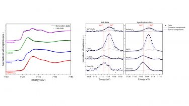
Good quality X-ray absorption or emission spectroscopy to be routinely acquired from materials with moderately dilute to concentrated absorbers in a matter of hours, including acquisition of the full Extended X-ray Absorption Fine Structure (EXAFS). Our benchmarking studies have demonstrated an effective resolution comparable to third generation synchrotron bending magnet beamlines utilising a Si (111) double crystal monochromator, in favourable circumstances. Importantly, this enables acquisition of X-ray Absorption Near Edge Structure (XANES), with sufficient resolution to permit determination of absorber speciation from the near edge and weak pre-edge features, as demonstrated by the Fe K-edge XANES data in Figure 1 [3]. Note that the Fe concentration was 4 mol. % in the staurolite reference compound reported in Figure 1, demonstrating the potential for rapid and routine Fe speciation by lab XANES even for moderately dilute absorber concentrations in transmission mode.
Figure 1: Comparison of lab and synchrotron XAS data at Fe K-edge for typical reference compounds, showing fit to weak pre-edge features for determination of speciation. Lab studies used Ge(620) monochromator; synchrotron data were acquired at the ESRF DUBBLE beamline (BM 26) with Si (111) channel cut monochromator. Data acquired in transmission mode with 0.25 eV step in XANES region for both studies [3].
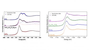
Additionally, we have shown lab XANES data to have sufficient resolution and reproducibility to enable routine analytical characterisation of lanthanide and actinide compounds to determine average oxidation state [4]. Figure 2 presents example lab XANES data acquired at the Ce L3 and U L3 edge, which demonstrate excellent agreement with synchrotron data and the feasibility of speciation by linear combination analysis or the chemical shift of the X-ray absorption edge. Note the application of these methods to complex materials with Ce and U concentration at realistic, but relatively dilute, concentration of a few mol.%, relevant to the ceramic immobilisation of actinides.
Figure 2: Comparison of lab and synchrotron XAS data at Ce L3 and U L3-edges for complex ceramics and reference compounds [4]. Data acquired in transmission mode with 0.75 eV or 0.5 eV step in XANES region for lab and synchrotron studies, respectively. Lab data were acquired with the (422) and (1266) harmonic of a Si (211) SBCA, at the Ce L3 and U L3-edge respectively. Synchrotron data were acquired at beamline B18, Diamond Light Source, configured with a fixed-exit double crystal Si(111) monochromator.
References
- G.T. Seidler, D.R. Mortensen, A.J. Remesnik, J.I. Pacold, N.A. Ball, N. Barry, M. Styczinski, O.R. Hoidn, A laboratory-based hard x-ray monochromator for high-resolution x-ray emission spectroscopy and x-ray absorption near edge structure measurements, Rev. Sci. Instrum., 2014, 85, 113906.
- E.P. Jahrman, W.M. Holden, A.S. Ditter, D.R. Mortensen, G.T. Seidler, T.T. Fister, S.A. Kozimor, L.F.J. Piper, J. Rana, N.C. Hyatt, M. Stennett, A laboratory-based hard x-ray monochromator for high-resolution x-ray emission spectroscopy and x-ray absorption near edge structure measurements, Rev. Sci. Instrum., 2019, 90, 024106.
- L.M. Mottram, S. Cafferkey, A.R. Mason, T. Oulton, S.K. Sun, D.J. Bailey, M.C. Stennett, N.C. Hyatt, A feasibility investigation of speciation by Fe K-edge XANES using a laboratory X-ray absorption spectrometer, J. Geosci., 2020, 65, 27-35.
- L.M. Mottram, M.C. Dixon Wilkins, L.R. Blackburn, T. Oulton, M.C. Stennett, S.K. Sun, C.L. Corkhill, N.C. Hyatt, A Feasibility Investigation of Laboratory Based X-ray Absorption Spectroscopy in Support of Nuclear Waste Management, MRS Adv., 2020, 5, 27-35.


